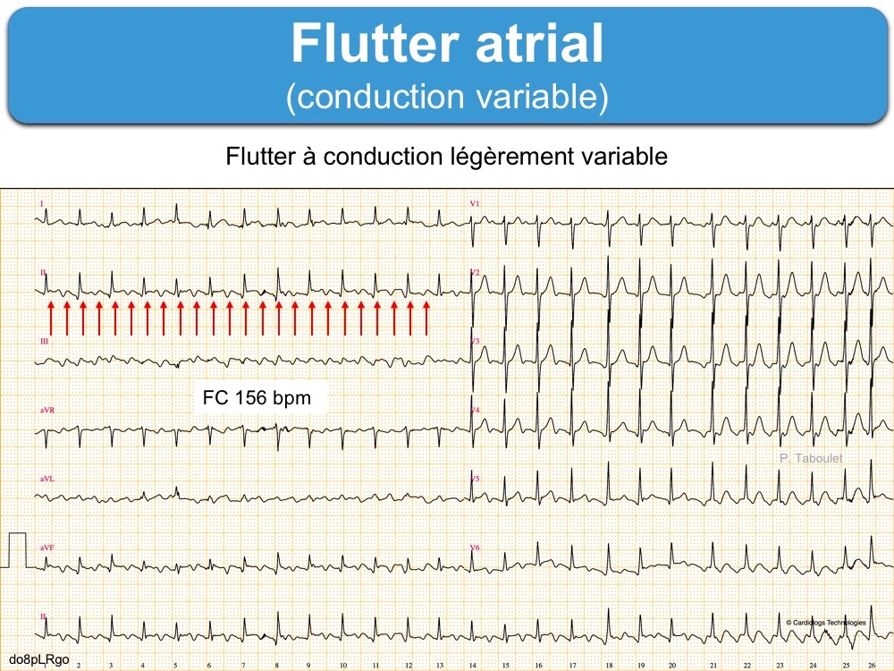


The mean left atrial pressure prior to the valvotomy was 31 mmHg. A transeptal puncture was performed after proper needle tip position was confirmed by fluoroscopy (right anterior oblique, left anterior oblique and 90° lateral views) and transoesophageal echocardiography (bicaval and short axis views Figure 3A). On the day of the procedure, the first balloon mitral valvotomy was performed from a right femoral approach using a 23–26 mm Accura balloon (Vascular Concepts) after transeptal access using an 8 Fr SL-1 sheath and a BRK-0 needle (St Jude Medical). Oral verapamil was initiated and the atrial flutter reverted to normal sinus rhythm. The patient’s blood pressure was 124/80 mmHg. A 12-lead ECG revealed atrial flutter with right bundle branch aberrancy on metoprolol succinate ( Figure 2B). The day before the procedure, the patient developed an episode of palpitation during the clinical round. Informed consent was obtained for the procedure. The coronary angiogram was normal.Īn electrophysiologist, cardiothoracic surgeon, cardiac anaesthetist and cardiologist suggested mitral valvotomy followed by ablation of the accessory pathway in a single procedure if possible to avoid repeated septal puncture. The pliable mitral valve area was 0.8 cm 2 and the mean gradient was 17 mmHg at a heart rate of 87 BPM. A transoesophageal echocardiogram confirmed rheumatic mitral stenosis ( Figure 2A and Supplementary Material Video 1). A chest X-ray in the posterior–anterior view was consistent with cardiac auscultation ( Figure 1B). A 12-lead ECG was suggestive of a WPW pattern with a right posterior septal accessory pathway ( Figure 1A). Cardiac auscultation raised the suspicion of underlying rheumatic mitral stenosis. Each episode spontaneously reverted to the normal sinus rhythm 6–8 hours after the onset. 5,6Ī 57-year-old man presented with a three-decade history of recurrent episodes of palpitation with alarming jugular venous pulsation, but without syncope. Among these, balloon mitral valvotomy and radiofrequency ablation were performed simultaneously in only two cases. 4 The coexistence of rheumatic mitral stenosis and WPW syndrome is so rare that a search of the literature using Google Scholar and PubMed yielded 17 cases between 19. 3 The prevalence of Wolff–Parkinson–White (WPW) syndrome or the WPW pattern in the general population ranges from 0.1 to 0.3%, with a higher prevalence among men than women. 1,2 However, based on echocardiography, the prevalence of rheumatic heart disease is likely to be greater. Providers should ensure that governance structures are in place to review, authorise and monitor the introduction of new devices and procedures.Ĭommissioners and providers have a responsibility to promote an environmentally sustainable health and care system and should assess and reduce the environmental impact of implementing NICE recommendations wherever possible.In India, the prevalence of rheumatic heart disease in the general population is 1.5–2.0 per 1,000. Nothing in this guidance should be interpreted in a way that would be inconsistent with compliance with those duties. The guidance does not override the individual responsibility of healthcare professionals to make decisions appropriate to the circumstances of the individual patient, in consultation with the patient and/or guardian or carer.Īll problems (adverse events) related to a medicine or medical device used for treatment or in a procedure should be reported to the Medicines and Healthcare products Regulatory Agency using the Yellow Card Scheme.Ĭommissioners and/or providers have a responsibility to implement the guidance, in their local context, in light of their duties to have due regard to the need to eliminate unlawful discrimination, advance equality of opportunity, and foster good relations. When exercising their judgement, healthcare professionals are expected to take this guidance fully into account, and specifically any special arrangements relating to the introduction of new interventional procedures. This guidance represents the view of NICE, arrived at after careful consideration of the evidence available.


 0 kommentar(er)
